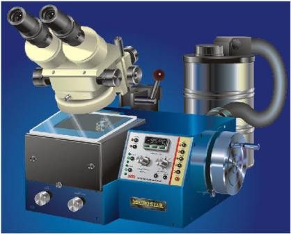
It’s cold. Very cold. And that’s exactly why it’s changing science.
Cryomicroscopy, or cryo-EM, is a technique that freezes samples so fast they don’t even realize they’re dying. Developed in 1978, it lets researchers look at macromolecules in their natural state. No dyes. No dehydration. Just pure, undisturbed structure.
The name comes from the Greek word for “cold.” That’s not a detail. It’s the entire point.
How Cryo-EM Works Without Destroying Your Sample
Traditional electron microscopy requires you to dehydrate or stain samples. That changes them. It kills the native structure. Cryo-EM skips that step.
Instead, it uses vitrification. The sample is plunged into liquid ethane or liquid nitrogen. The cooling is ultrarapid. It happens faster than water molecules can arrange themselves into ice crystals.
Ice crystals are the enemy. They puncture delicate biological structures. Vitrification creates a glass-like solid. The molecules stay exactly where they were. They are frozen in time.
“The ultra-fast cooling prevents the formation of ice crystals that would otherwise damage fine structures.”
The sample is mounted on special grids. It’s kept at around -180 °C throughout the observation. This stops sublimation. It stops degradation from the electron beam. You get high contrast images of proteins and nucleic acids without artificial contrast agents that could skew results.
It’s neutral. It’s invasive-free. It just watches.
Why Researchers Swapped X-Ray Crystals for Cryo-EM
For decades, scientists relied on X-ray crystallography to see molecules. If you could crystallize the target, you were good. If you couldn’t, you were stuck.
Many complex biological assemblies refuse to crystallize. Viruses. Ribosomes. Membrane proteins. They’re too floppy. Too dynamic.
Cryo-EM doesn’t need crystals. It looks at single particles. This is where the revolution happened.
The technique allows for single particle analysis. Computers reconstruct 3D structures from thousands of 2D images. The resolution now reaches the atomic level. You can see how proteins fold. You can see how they interact. You can see the shape of a virus envelope in exquisite detail.
This isn’t just incremental. It’s a paradigm shift.
Where Cryo-EM Is Being Used Today
Biologists aren’t the only ones excited. This isn’t just for life sciences.
Material science has jumped on board. Researchers use cryo-EM to study nanostructures. They look at polymers. They examine advanced materials under extreme conditions. It reveals internal organization and defects without altering the sample. It’s crucial for optimizing new devices.
Chemists use it to capture intermediate states. Reactions that happen too fast to see? Cryo-EM freezes them. It captures phase transitions in real-time. It shows what happens in the milliseconds between reactants and products.
Medicine is feeling the impact too. Cryo-tomography allows for the 3D reconstruction of whole cells. You get volumes. You get organelles. You get architecture described with a finesse never before possible.
This matters for neurodegenerative diseases. It matters for virology. It matters for drug design. If you can see the 3D shape of a molecular target precisely, you can design drugs that fit it like a key in a lock.
The Future Is Cold
The technology is still developing. The equipment is expensive. The expertise required is deep. But the payoff is undeniable.
We are no longer guessing at the shapes of biological machines. We are seeing them. In their native state. At atomic resolution.
As algorithms improve and detectors become more sensitive, the resolution will only get better. The questions we can ask will get sharper.
Why wait for crystals when you can just freeze the moment? The answer is simple. You don’t have to wait. You just have to get cold.
How cryo-EM moved from niche instrument to structural biology powerhouse
The shift in electron microscopy wasn’t gradual. It was a collision of hardware and software that finally broke the resolution barrier. Early cold-stage microscopes were stuck with blurry, low-res images. You couldn’t see the details. Then came direct electron detectors. These cameras don’t just record hits; they track individual electrons. Pair that with smarter image processing algorithms and full automation, and the entire field pivoted.
Now, researchers can map proteins and massive supramolecular assemblies at sub-nanometer resolution. This isn’t just a tweak. It’s a complete rewrite of how we understand molecular mechanics. If you’re looking into the future of cryo-EM resolution limits, the trend is clear: we are getting closer to atomic-level detail for increasingly complex biological machines.
Why sample prep remains the bottleneck
High resolution sounds great until you realize you have to feed the machine perfect samples. That’s where the friction lies. Getting homogeneous samples is hard. Background noise is everywhere. And the data output? It’s overwhelming.
Reconstructing 3D models from 2D projections is computationally expensive. It requires serious computing power and specialized skills. You can’t just walk in and run the software. The entire process—from freezing the sample to final analysis—demands a stable cryogenic environment. One fluctuation in temperature, and your grid is ruined.
Recent updates focus on robustness. Automated vitrification. Robotic grid preparation. AI-driven particle picking. These aren’t just buzzwords. They are solutions to reproducibility. When protocols become automated, they become scalable. This allows for large-scale, systematic analysis rather than one-off, painstaking experiments.
Multimodal imaging: Breaking down silos
The next big leap isn’t just better microscopes. It’s better integration. The current trajectory points toward multimodal approaches. Think cryo-EM combined with fluorescence microscopy. Or integrating spectroscopy. Even X-ray tomography.
Why combine them? Because structure alone doesn’t tell the whole story. Fluorescence shows you where things are in a cell. Spectroscopy tells you about chemical states. Cryo-EM gives you the atomic blueprint. Merging these methods cross-references structural and functional data. You get an integrated understanding of biological phenomena. This interdisciplinary mix is why there’s so much funding flowing into molecular biology, medicine, and materials science right now.
Institutional backing drives adoption
This technology isn’t floating in a vacuum. It’s anchored by major institutions. Platforms for integrated structural biology are becoming critical hubs. They develop the tech and disseminate it. The CNRS, for example, highlights the role of these dedicated platforms in advancing the field. Without this infrastructure, the complexity of cryo-EM would remain inaccessible to all but a handful of labs.